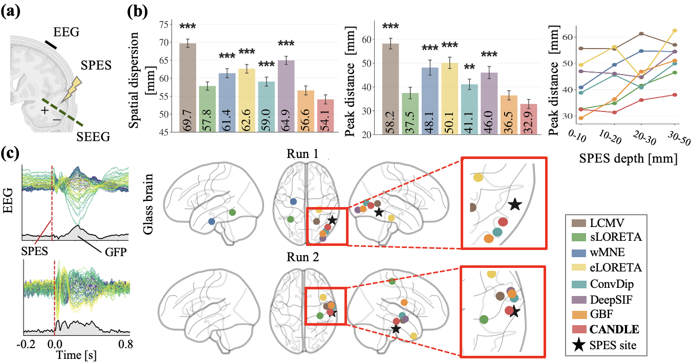
We proposed CANDLE, a learning-based electrophysiological source imaging model that estimates source activity on subject-specific cortical geometries. CANDLE learns subject-specific null-space priors from large-scale simulations and generalizes zero-shot to empirical tasks. Here are some demo videos of CANDLE for intracranial stimulation localization.
 CANDLE
Cortical Null-Space Decomposition
for Noninvasive Brain Source Imaging
CANDLE
Cortical Null-Space Decomposition
for Noninvasive Brain Source Imaging
Under review
TL;DR
Abstract
Electrophysiological source imaging (ESI) aims to estimate cortical source activity from noninvasive electrophysiological measurements such as electroencephalogram (EEG). However, ESI is fundamentally ill-posed because source activity is substantially higher-dimensional than sensor observations, resulting in non-unique solutions. Recent learning-based approaches address this ambiguity by learning data-driven source priors, yet they often struggle to generalize across subject-specific cortical geometries.
To address this, we propose CANDLE, a learning-based ESI model that estimates source activity on subject-specific cortical geometries. CANDLE learns a prior over the null space induced by the source-to-sensor mapping derived from T1-weighted MRI, restricting learning to unobservable source components while preserving geometric constraints. To train CANDLE, we develop a whole-brain simulator spanning over 1,100 subject-specific cortical geometries with source configurations derived from over 26,000 statistical brain maps.
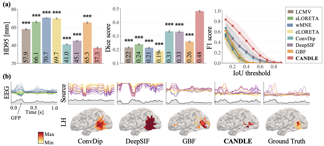
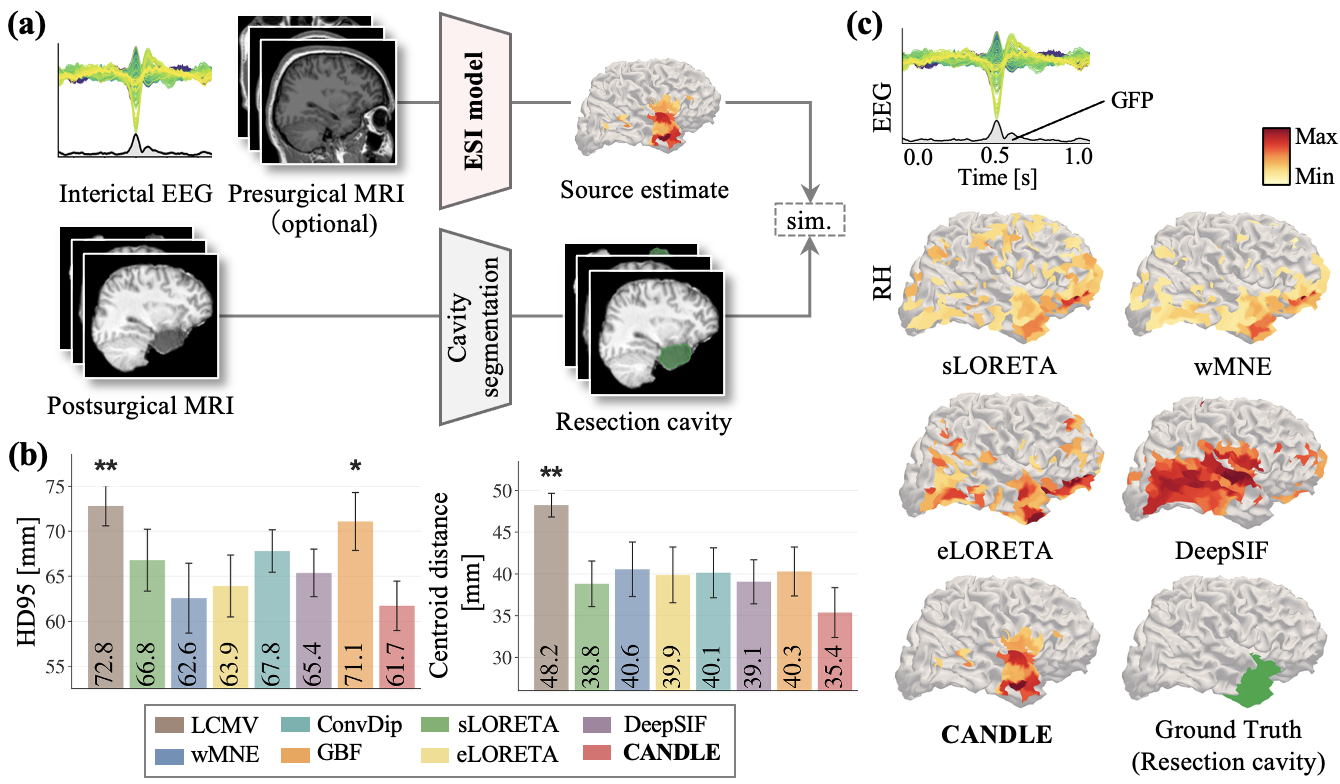
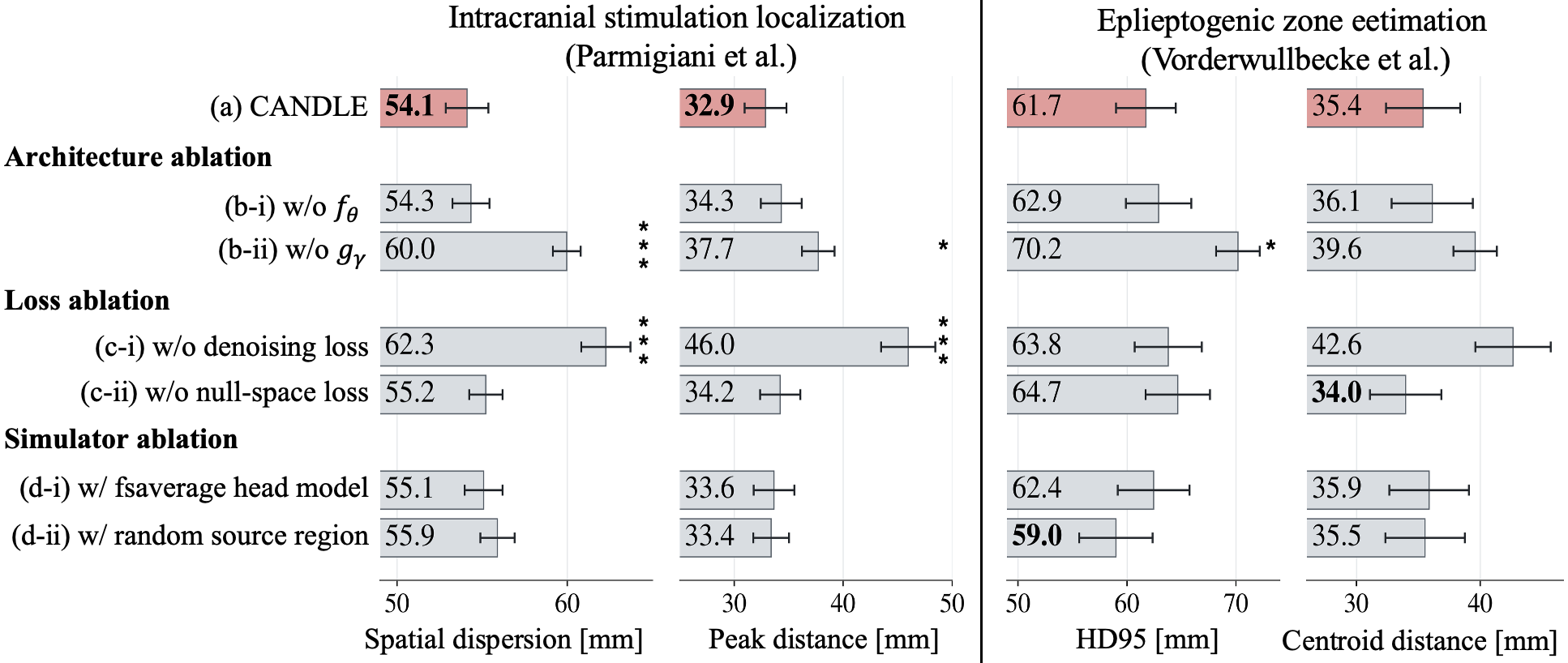
Trained exclusively on simulated data, CANDLE outperformed prior ESI methods on simulated source activity estimation and generalized to two empirical tasks: (i) intracranial stimulation localization from simultaneously recorded scalp EEG and (ii) epileptogenic zone estimation from presurgical interictal EEG.
Method
1. CANDLE architecture
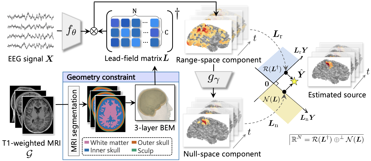
CANDLE derives a lead-field matrix \(\mathbf{L}\) from each subject's T1-weighted MRI. The null-space projector \(\mathbf{L}_{\mathrm{n}} = \mathbf{I} - \mathbf{L}^{\dagger}\mathbf{L}\) isolates the component of source activity that is unobservable from EEG. After denoising the EEG with \(f_\theta\), \(\mathbf{L}^{\dagger}\mathbf{X}_{\mathrm{denoise}}\) analytically recovers the observable component of source activity (i.e., the range-space component). Meanwhile, \(g_\gamma\) predicts a plausible source distribution from the observable component, and \(\mathbf{L}_{\mathrm{n}}\) extracts the unobservable component (i.e., the null-space component) of this prediction. The observable and predicted null-space components are then added together, preserving consistency with the denoised EEG. The final source estimate \(\hat{\mathbf{Y}}\) is obtained as follows:
\[
\begin{aligned}
\mathbf{X}_{\mathrm{denoise}} &= f_{\theta}(\mathbf{X}), \\
\hat{\mathbf{Y}} &=
\underbrace{\mathbf{L}^{\dagger}\mathbf{X}_{\mathrm{denoise}}}_{\text{Range-space component}}
+
\underbrace{\mathbf{L}_{n}g_{\gamma}\!\left(\mathbf{L}^{\dagger}\mathbf{X}_{\mathrm{denoise}}\right)}_{\text{Null-space
component}}.
\end{aligned}
\]
2. Training CANDLE
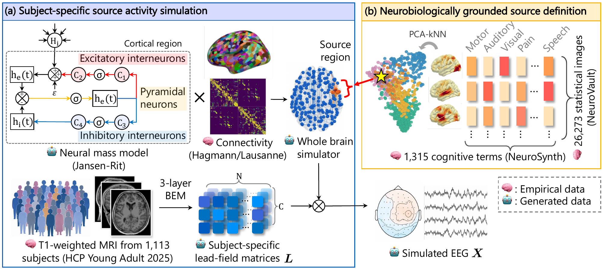
CANDLE is trained entirely on synthetic data from a whole-brain simulator spanning 1,113 subject-specific cortical geometries. Coupled Jansen–Rit neural mass models generate cortical dynamics, while source configurations derived from 26,273 statistical brain maps provide diverse, neurobiologically grounded activity patterns that are projected to EEG through each subject’s lead-field matrix.
Results
BibTeX
Coming soon...